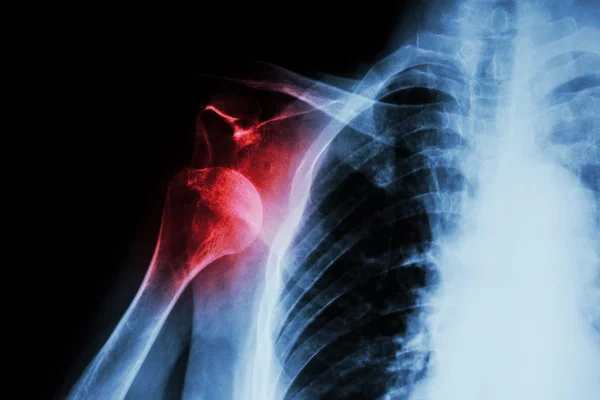
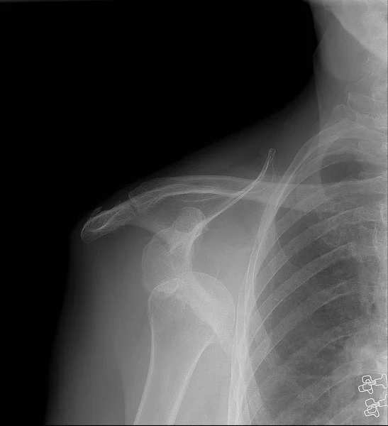
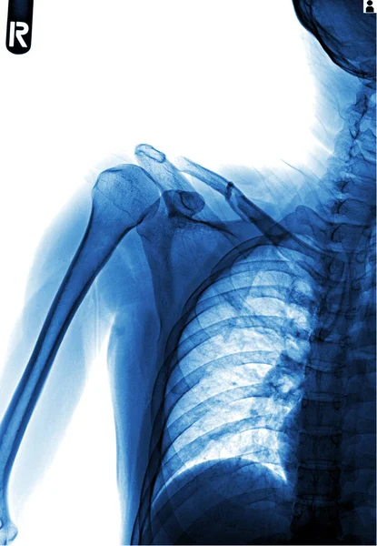
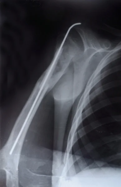
X-ray anterior shoulder dislocation
X-ray anterior shoulder dislocation — Stock Photo, Image
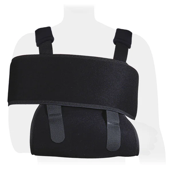
Arm Brace Shoulder Brace
Arm Brace Shoulder Brace — Stock Photo, Image
Orthopedist Massaging the shoulder of a Female Patient
Orthopedist Massaging the shoulder of a Female Patient — Stock Photo, Image
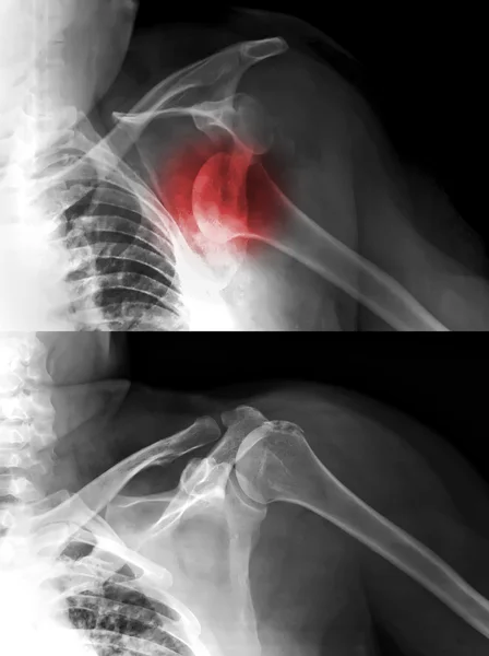
X-rays image of shoulder dislocation
X-rays image of shoulder dislocation — Stock Photo, Image
X-ray anterior shoulder dislocation
X-ray anterior shoulder dislocation — Stock Photo, Image
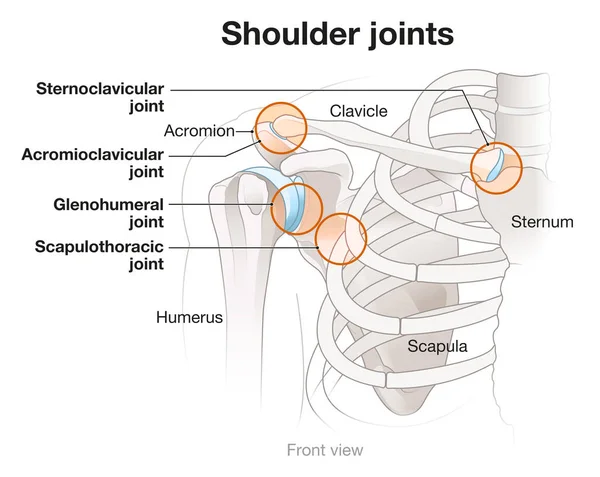
Shoulder anatomy. Four major joints of the shoulder.
Shoulder Anatomy Four Major Joints Shoulder — Stock Photo, Image
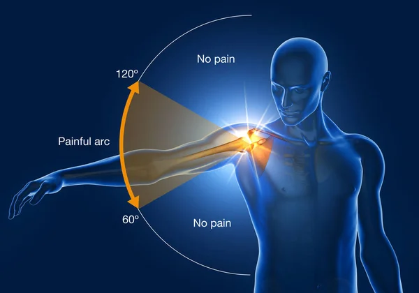
Illustration showing shoulder impingement, painfull arc, 3D illustration
Illustration Showing Shoulder Impingement Painfull Arc Illustration — Stock Photo, Image
Bearded man touching shoulder with hand pain dislocation medical intervention
Bearded man touching shoulder with hand pain dislocation medical intervention — Stock Photo, Image
X-ray anterior shoulder dislocation
X-ray anterior shoulder dislocation — Stock Photo, Image
Man with pain in shoulder. Pain in the human body
Man with pain in shoulder. Pain in the human body — Stock Photo, Image
Arm Brace Shoulder Brace
Arm Brace Shoulder Brace — Stock Photo, Image
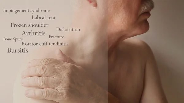
Shoulder pain words on senior man
Shoulder pain words on senior man — Stock Photo, Image
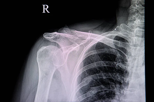
X-ray image of shoulder, Shows right shoulder dislocation. Anterior projection
X-ray image of shoulder, Shows right shoulder dislocation. Anterior projection — Stock Photo, Image
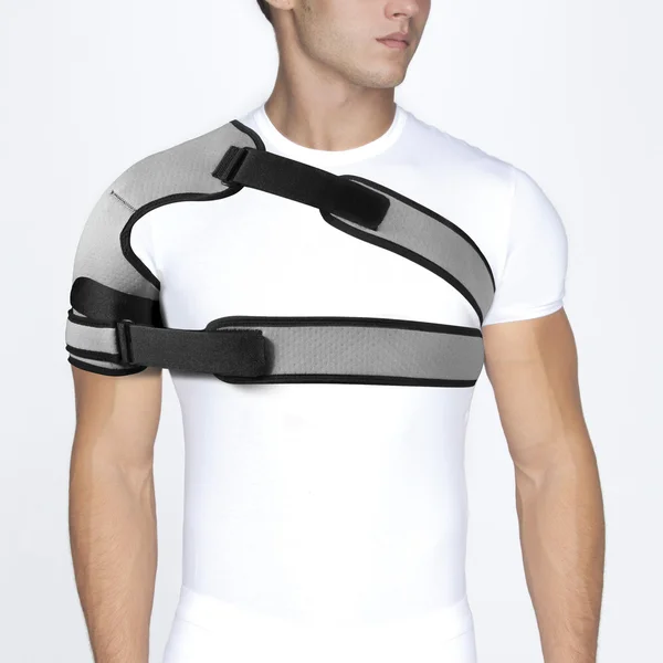
Shoulder Joint Brace, shoulder fixation
Shoulder Joint Brace, shoulder fixation — Stock Photo, Image
Man showing pain concept
Man showing pain concept — Stock Photo, Image
Man suffering from neck or shoulder pain
Man suffering from neck or shoulder pain — Stock Photo, Image
Shoulder Joint Brace, shoulder fixation
Shoulder Joint Brace, shoulder fixation — Stock Photo, Image
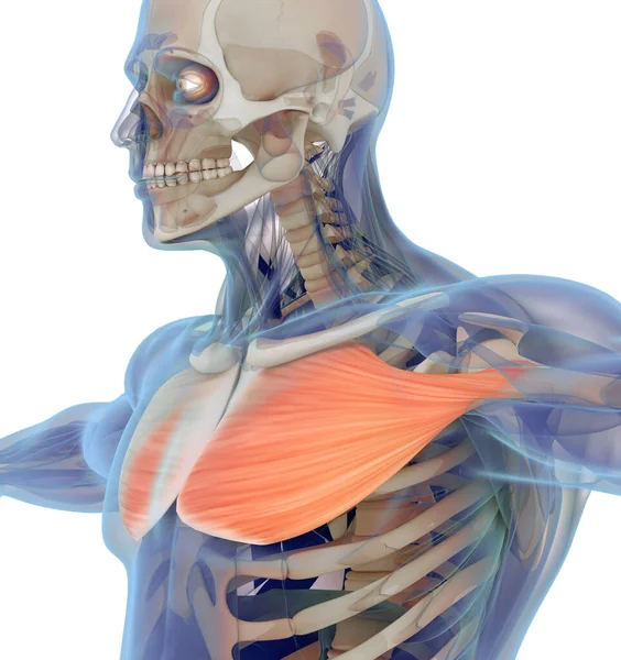
Medical muscle illustration of the pectoralis major. 3d illustration
Medical Muscle Illustration Pectoralis Major Illustration — Stock Photo, Image
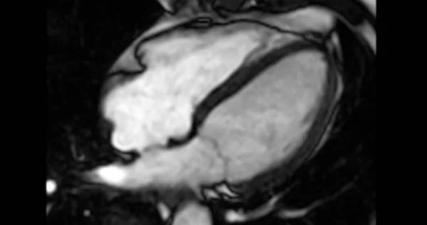
MRI heart or Cardiac MRI ( magnetic resonance imaging ) of heart in Short axis view showing heart beating of 4 chamber for detecting heart disease.
Mri Heart Cardiac Mri Magnetic Resonance Imaging Heart Short Axis — Stock Photo, Image
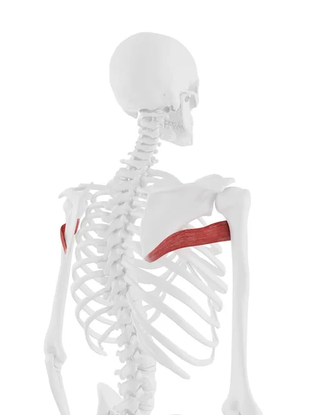
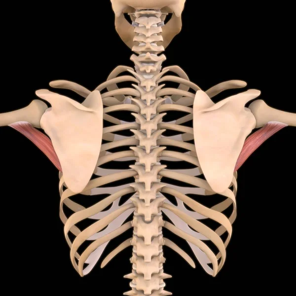
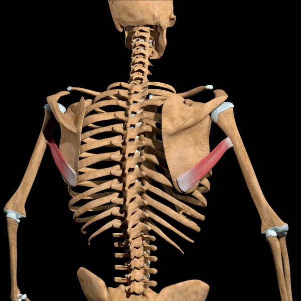
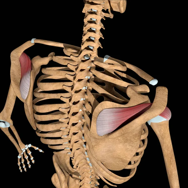
Human skeleton model with detailed Teres major muscle, computer illustration.
Human Skeleton Model Detailed Teres Major Muscle Computer Illustration — Stock Photo, Image
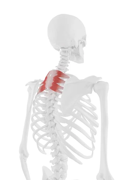
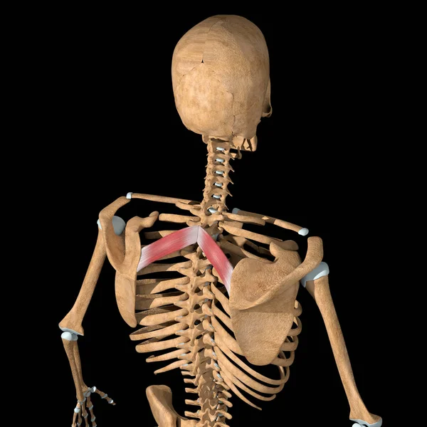
Human skeleton with red colored Serratus posterior superior muscle, digital illustration.
Human Skeleton Red Colored Serratus Posterior Superior Muscle Digital Illustration — Stock Photo, Image
X - ray of the human chest, which is an x - ray.
Ray Human Chest Which Ray — Stock Photo, Image
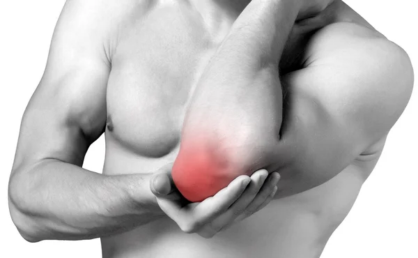
Strong man with cubit pain
Strong man with cubit  pain — Stock Photo, Image
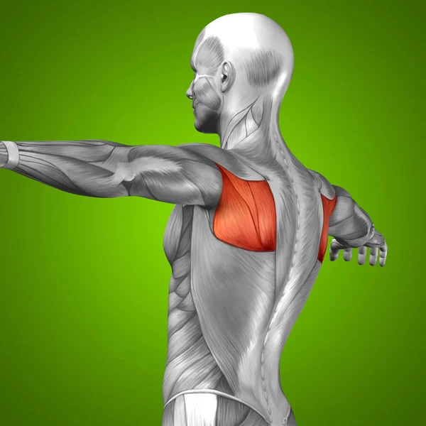
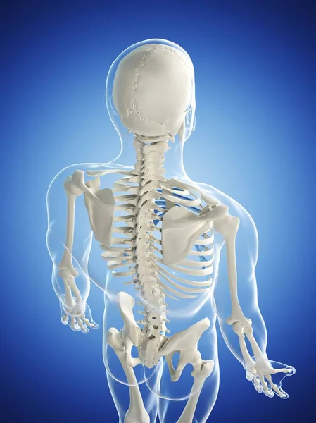
Back human anatomy
Back human anatomy — Stock Photo, Image
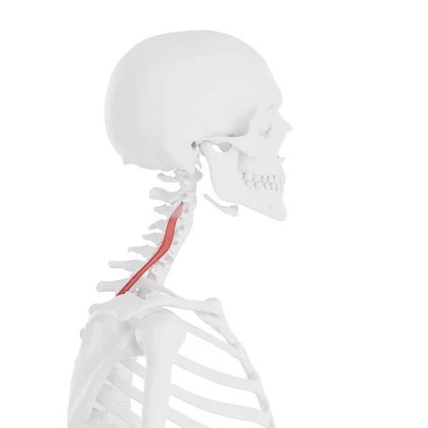
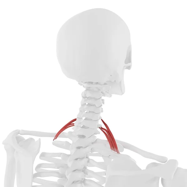
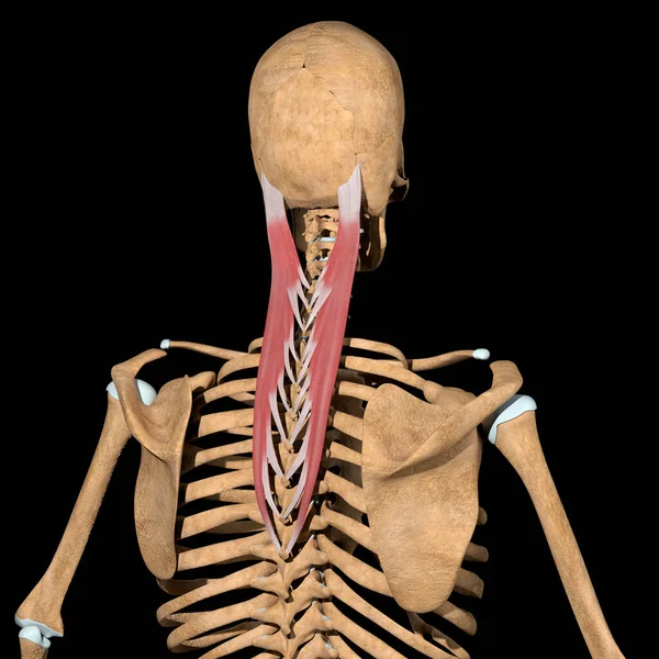
Human skeleton with red colored Longissimus cervicis muscle, digital illustration.
Human Skeleton Red Colored Longissimus Cervicis Muscle Digital Illustration — Stock Photo, Image
3D Illustration, Muscle is a soft tissue, Muscle cells contain proteins , producing a contraction that changes both the length and the shape of the cell. Muscles function to produce force and motion.
Illustration Muscle Soft Tissue Muscle Cells Contain Proteins Producing Contraction — Stock Photo, Image
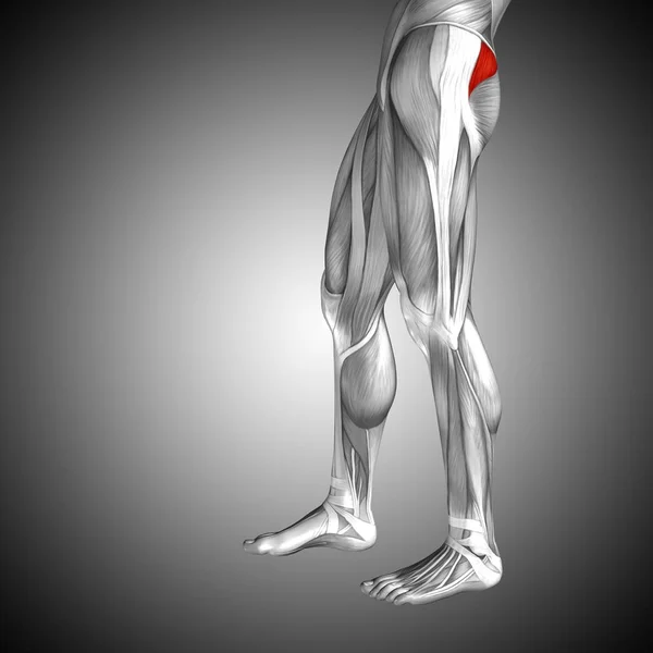
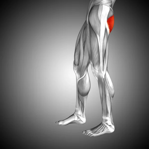
Human upper leg anatomy
Human upper leg anatomy — Stock Photo, Image
Arm Muscle Anatomy For Medical Concept 3D Illustration
Arm Muscle Anatomy Medical Concept Illustration — Stock Photo, Image
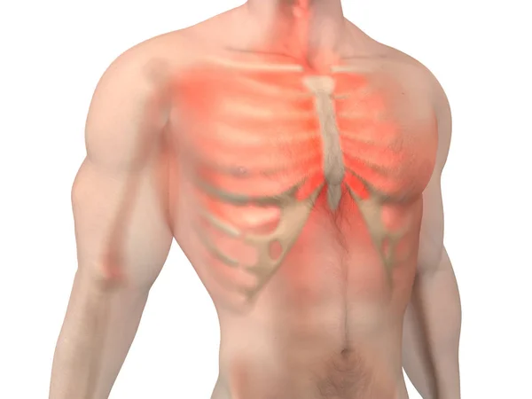
Male Anatomy - Chest Pain
Male Anatomy - Chest Pain — Stock Photo, Image
This 3d illustration shows the sternocleidomastoid muscles on white background
Illustration Shows Sternocleidomastoid Muscles White Background — Stock Photo, Image
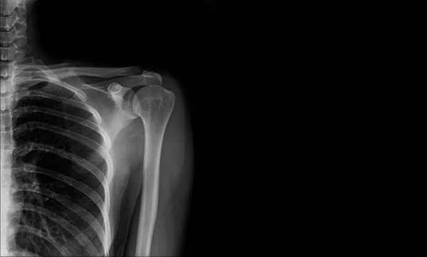
X-ray shoulder banner for website or work print
Ray Shoulder Banner Website Work Print — Stock Photo, Image
Верхней части плечевой кости
Верхней части плечевой кости — Stock Photo, Image
3D Illustration, Muscle is a soft tissue, Muscle cells contain proteins , producing a contraction that changes both the length and the shape of the cell. Muscles function to produce force and motion.
Illustration Muscle Soft Tissue Muscle Cells Contain Proteins Producing Contraction — Stock Photo, Image
Male anatomy showing Deltoid muscle, computer illustration.
Male Anatomy Showing Deltoid Muscle Computer Illustration — Stock Photo, Image
Human skeleton with red colored Scalene posterior muscle, digital illustration.
Human Skeleton Red Colored Scalene Posterior Muscle Digital Illustration — Stock Photo, Image
Spine vertebra ribs back injury xray scan
Spine vertebra ribs back injury xray scan — Stock Photo, Image
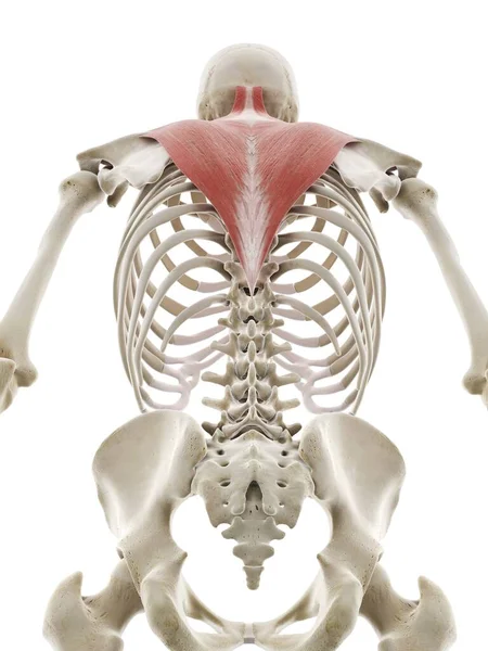
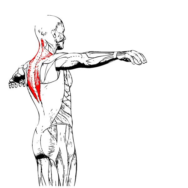
Trapezius muscle, computer illustration
Trapezius Muscle Computer Illustration — Stock Photo, Image
3D Illustration, Muscle is a soft tissue, Muscle cells contain proteins , producing a contraction that changes both the length and the shape of the cell. Muscles function to produce force and motion.
Illustration Muscle Soft Tissue Muscle Cells Contain Proteins Producing Contraction — Stock Photo, Image
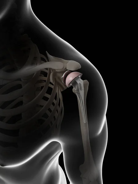
3d rendered illustration of a shoulder replacement
Rendered Illustration Shoulder Replacement — Stock Photo, Image
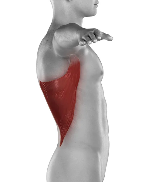
Man latissimus dorsi muscle
Man latissimus dorsi muscle — Stock Photo, Image
A chest xray of a patient with old ribs 5-7 fracture.
Chest Xray Patient Old Ribs Fracture — Stock Photo, Image
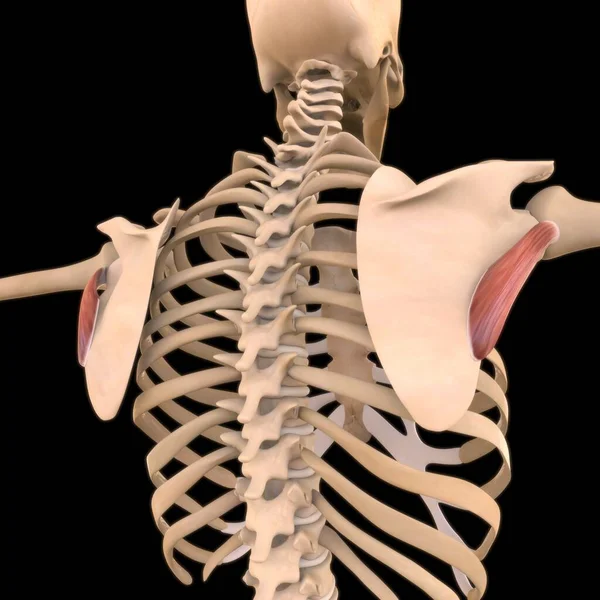
This 3d illustration shows the teres major muscles on skeleton
Illustration Shows Teres Major Muscles Skeleton — Stock Photo, Image
Coracobrachialis muscle. The deep muscles of the arm. Image on a black background.
Coracobrachialis muscle. The deep muscles of the arm. Image on a black background. — Stock Photo, Image
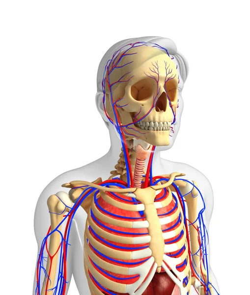
Male skeletal and circulatory system
Male skeletal and circulatory system — Stock Photo, Image
MRI heart or Cardiac MRI ( magnetic resonance imaging ) of heart in Short axis view showing heart beating of 4 chamber for detecting heart disease.
Mri Heart Cardiac Mri Magnetic Resonance Imaging Heart Short Axis — Stock Photo, Image
Bones of the neck and shoulder
Bones of the neck and shoulder — Stock Photo, Image
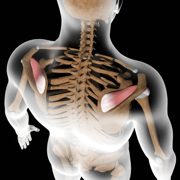
This 3d illustration shows the infraspinatus muscles on xray body
Illustration Shows Infraspinatus Muscles Xray Body — Stock Photo, Image
Shoulder blades and bones of shoulder girdle
Shoulder blades and bones of shoulder girdle — Stock Photo, Image
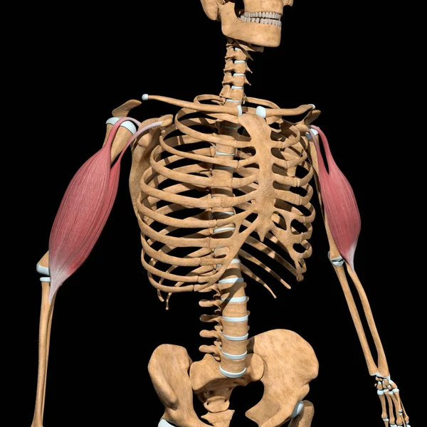
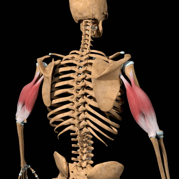
This 3d illustration shows the biceps muscles on skeleton
Illustration Shows Biceps Muscles Skeleton — Stock Photo, Image
Illustration of human body anatomy
Illustration of human body anatomy — Stock Photo, Image
Human skeleton with red colored Levator scapularis muscle, computer illustration.
Human Skeleton Red Colored Levator Scapularis Muscle Computer Illustration — Stock Photo, Image
Back human anatomy
Back human anatomy — Stock Photo, Image
Human upper leg anatomy
Human upper leg anatomy — Stock Photo, Image
Cervical spine vertebrae
Cervical spine vertebrae — Stock Photo, Image
Abdomen human anatomy
Abdomen human anatomy — Stock Photo, Image
3d rendered illustration of a shoulder replacement
Rendered Illustration Shoulder Replacement — Stock Photo, Image
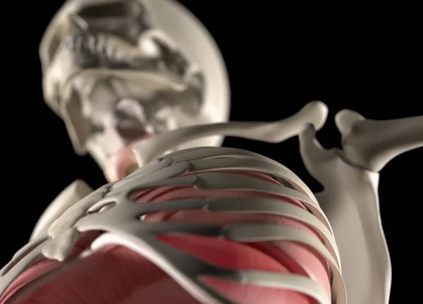
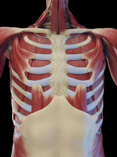
Human rib cage anatomy model
Human rib cage anatomy model — Stock Photo, Image
Brown ink stains texture, abstract background
Brown Ink Stains Texture Abstract Background — Stock Photo, Image
Back human anatomy
Back human anatomy — Stock Photo, Image
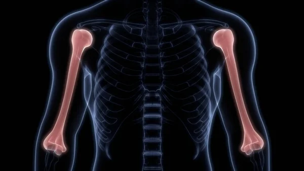
Humerus Bone Joints of Human Skeleton System Anatomy 3d rendering
Humerus Bone Joints Human Skeleton System Anatomy Rendering — Stock Photo, Image
X-ray of the human chest, the concept of preventive examination, treatment of injuries, lung diseases
Ray Human Chest Concept Preventive Examination Treatment Injuries Lung Diseases — Stock Photo, Image
A woman feeling exhausted and suffering from neck and head pain and injury on isolated white background. Health care and medical concept.
Woman Feeling Exhausted Suffering Neck Head Pain Injury Isolated White — Stock Photo, Image
Human skeleton with detailed red Middle scalene muscle, digital illustration.
Human Skeleton Detailed Red Middle Scalene Muscle Digital Illustration — Stock Photo, Image
Male deep torso muscles, illustration
Male Deep Torso Muscles Illustration — Stock Photo, Image
Biceps brachii Muscles
Biceps brachii Muscles — Stock Photo, Image
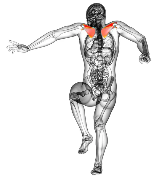
Human skeleton with red colored Trapezius muscle, computer illustration.
Human Skeleton Red Colored Trapezius Muscle Computer Illustration — Stock Photo, Image
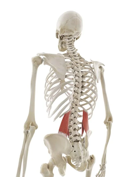
Quadratus lumborum muscle, computer illustration
Quadratus Lumborum Muscle Computer Illustration — Stock Photo, Image
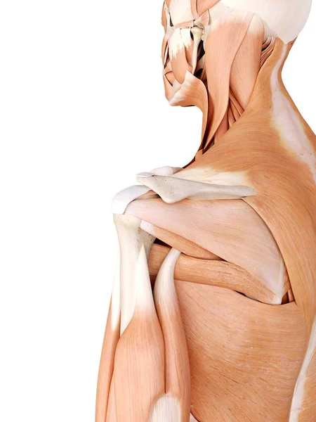
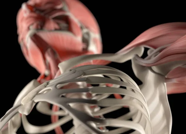
Shoulder musculature and structural anatomy
Shoulder musculature and structural anatomy — Stock Photo, Image
Human lungs and rib cage
Human lungs and rib cage — Stock Photo, Image
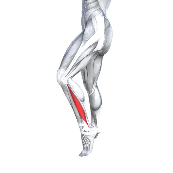
Concept conceptual 3D illustration fit strong front lower leg human anatomy, anatomical muscle isolated white background for body medical health tendon foot and biological gym fitness muscular system
Concept Conceptual Illustration Fit Strong Front Lower Leg Human Anatomy — Stock Photo, Image
Gray liquid ink, digital wallpaper
Gray Liquid Ink Digital Wallpaper — Stock Photo, Image
3d render medical illustration of the bladder
3d render medical illustration of the bladder — Stock Photo, Image
Back human anatomy
Back human anatomy — Stock Photo, Image
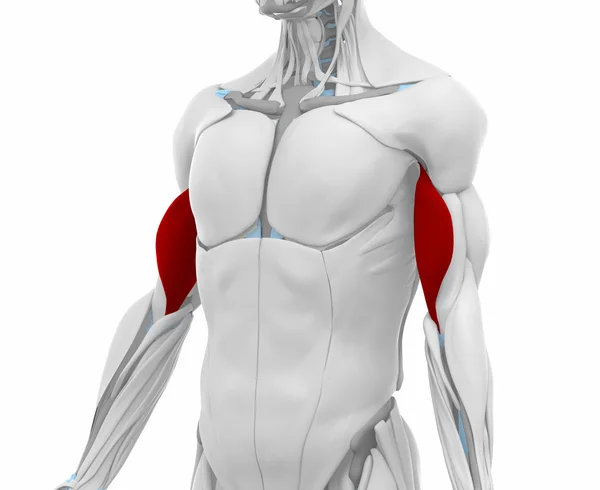
This 3d illustration shows the triceps muscles on skeleton
Illustration Shows Triceps Muscles Skeleton — Stock Photo, Image
Human chest anatomy
Human chest anatomy — Stock Photo, Image
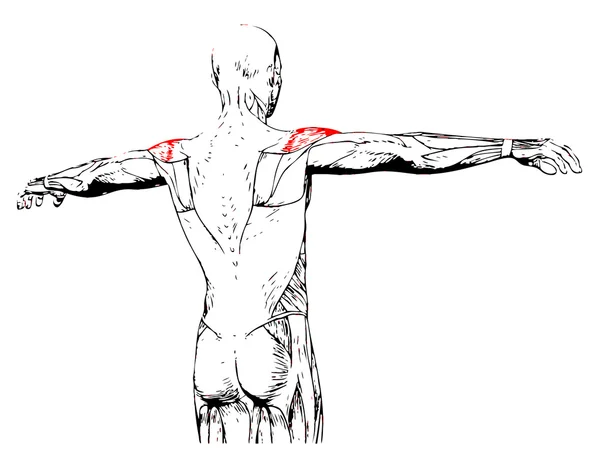
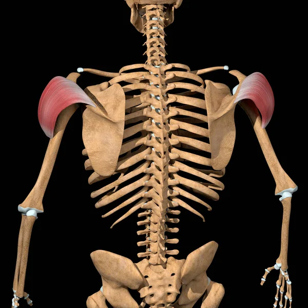
This 3d illustration shows the back view of the posterior head of the deltoid muscles
Illustration Shows Back View Posterior Head Deltoid Muscles — Stock Photo, Image
Therapist doing healing treatment on man's hand / shoulder. Alternative medicine, pain relief concept
Therapist doing healing treatment on man's hand / shoulder. Alternative medicine, pain relief concept — Stock Photo, Image
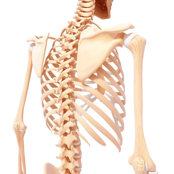
Bones of the back and shoulder blades
Bones of the back and shoulder blades — Stock Photo, Image
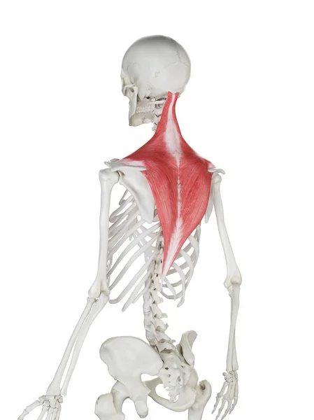
Human skeleton with red colored Trapezius muscle, computer illustration.
Human Skeleton Red Colored Trapezius Muscle Computer Illustration — Stock Photo, Image
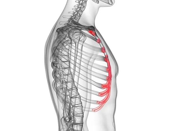
Abstract male silhouette with visible costal cartilage, computer illustration.
Abstract Male Silhouette Visible Costal Cartilage Computer Illustration — Stock Photo, Image
American football player suffering from neck pain
American football player suffering from neck pain — Stock Photo, Image
This 3d illustration shows the splenius capitis muscles on skeleton
Illustration Shows Splenius Capitis Muscles Skeleton — Stock Photo, Image
Human shoulder anatomy model
Human shoulder anatomy model — Stock Photo, Image
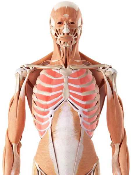
Chest and upper body muscles
Chest and upper body muscles — Stock Photo, Image
Human rib cage anatomy model
Human rib cage anatomy model — Stock Photo, Image
Shoulder girdle musculature
Shoulder girdle musculature — Stock Photo, Image
Backache
Backache — Stock Photo, Image
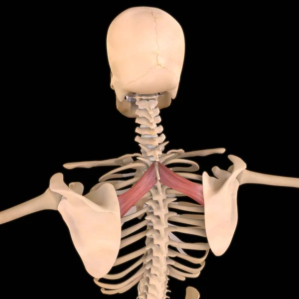
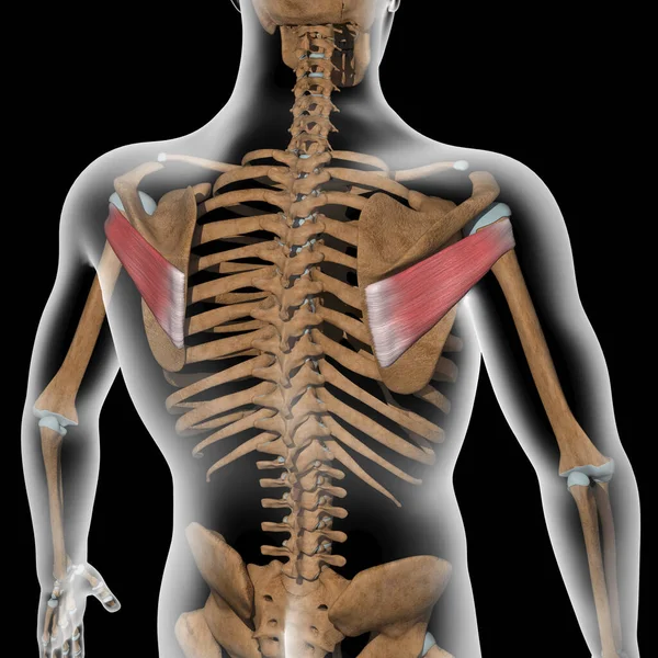
This 3d illustration shows the rhomboid minor muscles on skeleton
Illustration Shows Rhomboid Minor Muscles Skeleton — Stock Photo, Image
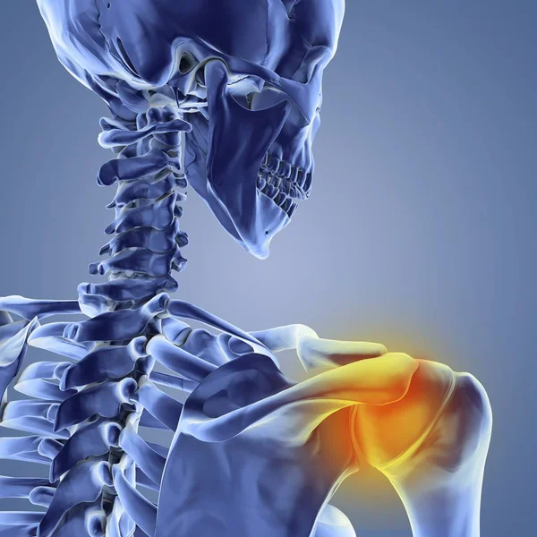
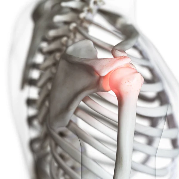
Digital illustration of painful shoulder joint in human skeleton.
Digital Illustration Painful Shoulder Joint Human Skeleton — Stock Photo, Image
This 3d illustration shows the supraspinatus muscles on xray body
Illustration Shows Supraspinatus Muscles Xray Body — Stock Photo, Image
Young Man Suffering From Elbow Pain
Young Man Suffering From Elbow Pain — Stock Photo, Image
3D Illustration, Muscle is a soft tissue, Muscle cells contain proteins , producing a contraction that changes both the length and the shape of the cell. Muscles function to produce force and motion.
Illustration Muscle Soft Tissue Muscle Cells Contain Proteins Producing Contraction — Stock Photo, Image
Human back anatomy
Human back anatomy — Stock Photo, Image
Shoulder joint bone structure
Shoulder joint bone structure — Stock Photo, Image
This 3d illustration shows the supraspinatus muscles on skeleton
Illustration Shows Supraspinatus Muscles Skeleton — Stock Photo, Image
3d render medical illustration of the scapula bone
3d render medical illustration of the scapula bone — Stock Photo, Image
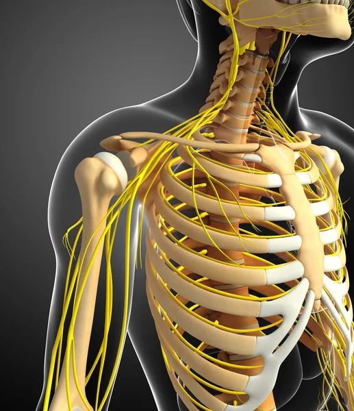
Nervous system and male ribcage artwork
Nervous system and male ribcage artwork — Stock Photo, Image

Shoulder Dislocation Images: Find the Perfect Visuals for Your Project

The Importance of Choosing the Right Images

When it comes to visual communication, images are worth a thousand words. That's why selecting the right visuals is crucial to make your message impactful and memorable. Whether you're creating educational materials, medical presentations, or advertising campaigns, including high-quality shoulder dislocation images is essential to convey your ideas effectively.

Our Stock Image Selection

At our stock image website, we offer a wide variety of shoulder dislocation images that come in JPEG, AI, and EPS formats. Our collection includes accurate and high-resolution photos, illustrations, and diagrams that illustrate different types of shoulder dislocations, such as anterior, posterior, and inferior. Besides, we also provide images that depict pain, swelling, bruising, and other symptoms related to shoulder dislocation.

Where and How to Use Our Images

Our shoulder dislocation images are perfect for various applications, such as medical textbooks, journal articles, online blogs, PowerPoint presentations, posters, flyers, and brochures. Depending on your project's purpose and audience, you can select images that cater to your visual style, color scheme, and tone of voice. For instance, if you're creating a medical presentation for a professional audience, you may prefer using detailed illustrations that highlight anatomical structures and surgical procedures. On the other hand, if you're creating a patient information brochure, you may opt for photos that depict real-life situations and emotions.

Maximizing the Impact of Shoulder Dislocation Images

To get the most out of our shoulder dislocation images, you should follow some best practices to make them more effective in your project. Firstly, always make sure the images are relevant to your message and create a clear link between the visual and text content. Secondly, choose images that are of high quality, resolution, and size so that they can be displayed correctly on different platforms and devices. Thirdly, consider adding captions, labels, and annotations that explain the context, source, and copyright information of the images. Finally, don't forget to test different variations of the images and evaluate their impact on your audience. With our shoulder dislocation images and these best practices, you can create remarkable visuals that resonate with your target audience and convey your message in a compelling way.