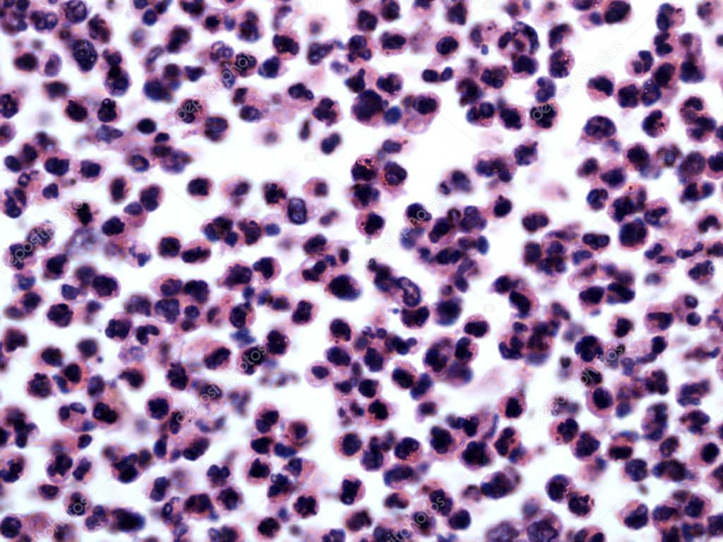
White blood cells of a human — Stock Photo, Image

White blood cells of a human — Photo

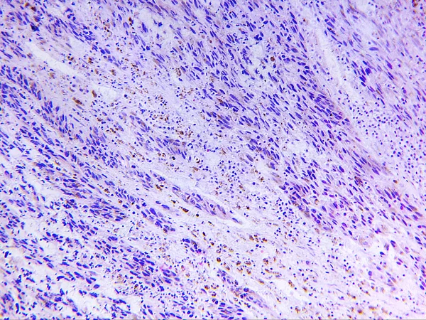
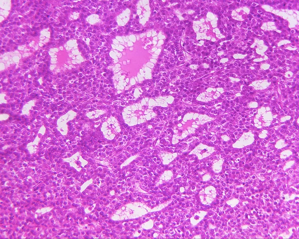
White blood cells of a human, photomicrograph panorama as seen under the microscope, 1000x zoom.

 — Photo by bond80

Same Series:

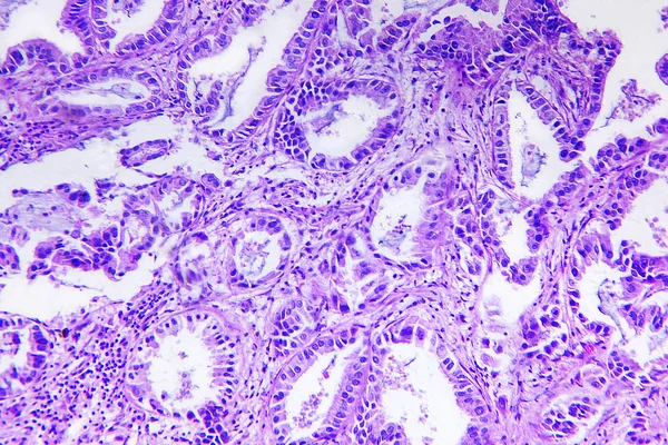
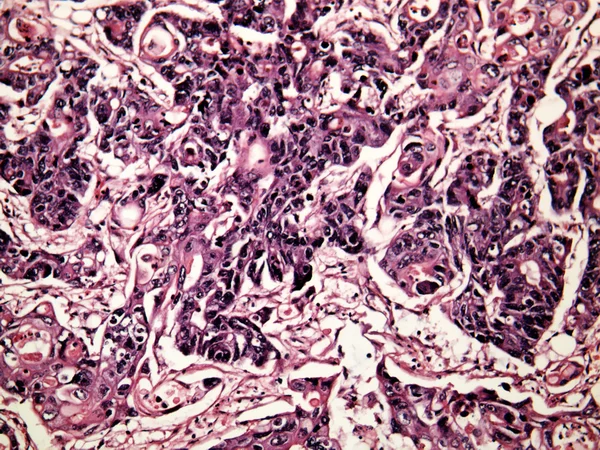
Well differentiated adenocarcinoma of a human, photomicrograph panorama as seen under the microscope, 200x zoom.
Well differentiated adenocarcinoma of a human — Stock Photo, Image
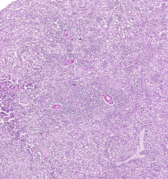
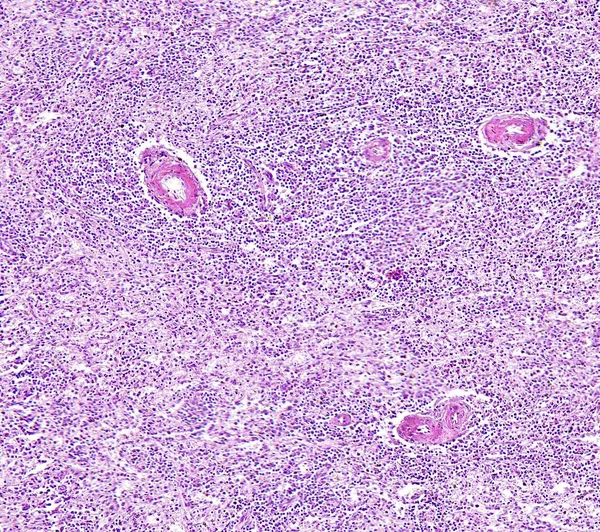
Central artery hyaline degeneration of spleen of a human, highly detailed segment of panorama. Photomicrograph as seen under the microscope, 10x zoom.
Central artery hyaline degeneration of spleen of a human — Stock Photo, Image
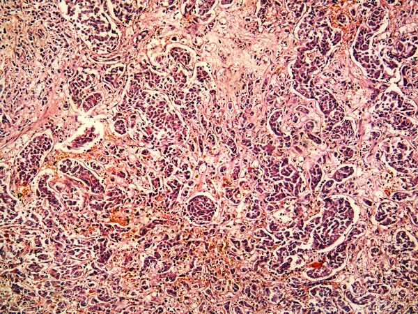
Melanoma of a human, photomicrograph panorama as seen under the microscope, 200x zoom.
Melanoma of a human — Stock Photo, Image
Hepatocellular carcinoma of a human, photomicrograph panorama as seen under the microscope, 200x zoom.
Hepatocellular carcinoma of a human — Stock Photo, Image
Histology of human tissue, show central artery hyaline degeneration of spleen as seen under the microscope
Histology of human tissue, show central artery hyaline degeneration of spleen as seen under the microscope, 10x zoom — Stock Photo, Image
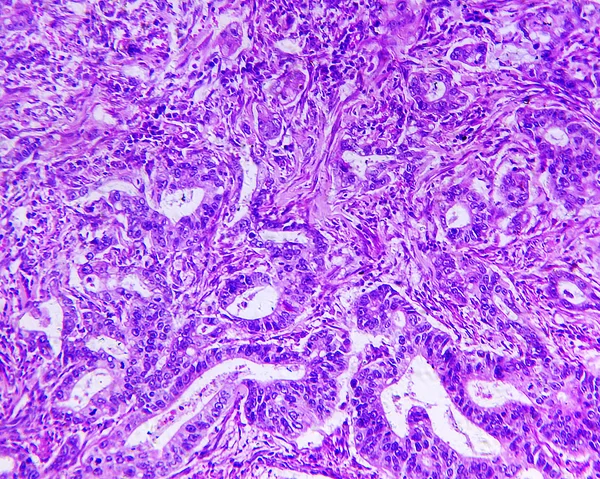
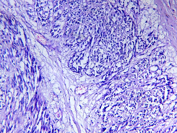
Gastric adenocarcinoma, tubular adenocarcinoma of a human, photomicrograph panorama as seen under the microscope, 200x zoom.
Gastric adenocarcinoma of a human — Stock Photo, Image
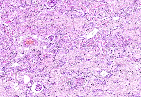
Histology of human tissue, show kidney proximal tubule epithelial cloudy swelling as seen under the microscope
Histology of human tissue, show kidney proximal tubule epithelial cloudy swelling as seen under the microscope, 10x zoom — Stock Photo, Image
Hepatocellular cancer of liver of a human, metastasises in vessels, photomicrograph panorama as seen under the microscope, 100x zoom.
Hepatocellular cancer of liver of a human — Stock Photo, Image
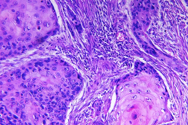
Squamous cell carcinoma of a human, photomicrograph panorama as seen under the microscope, 200x zoom.
Squamous cell carcinoma of a human — Stock Photo, Image
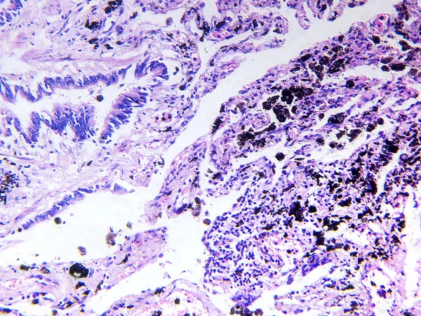
Lung of smoking, photomicrograph panorama as seen under the microscope, 200x zoom.
Lung of smoking — Stock Photo, Image
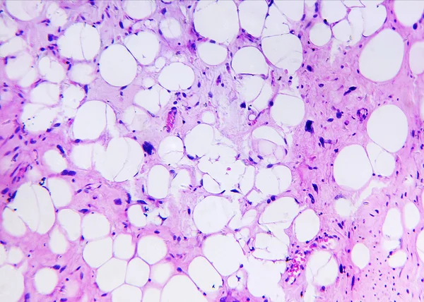
Liposarcoma of a human, photomicrograph panorama as seen under the microscope, 200x zoom.
Liposarcoma of a human — Stock Photo, Image
Histology of human tissue, show cortical necrosis of kidney as seen under the microscope
Histology of human tissue, show cortical necrosis of kidney as seen under the microscope, 10x zoom — Stock Photo, Image
Cortical necrosis of kidney of a human, highly detailed segment of panorama. Photomicrograph as seen under the microscope, 10x zoom.
Cortical necrosis of kidney of a human — Stock Photo, Image
Leiomyosarcoma of a human, photomicrograph panorama as seen under the microscope, 200x zoom.
Leiomyosarcoma of a human — Stock Photo, Image
Liver cancer of a human, photomicrograph panorama as seen under the microscope, 200x zoom.
Liver cancer of a human — Stock Photo, Image
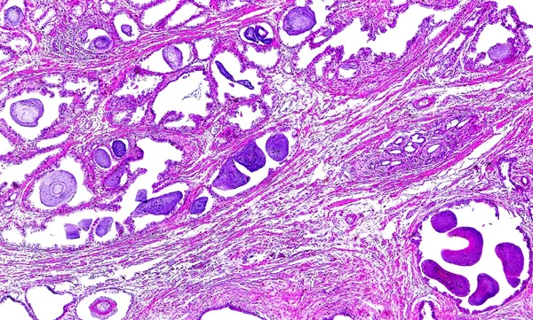
Prostate hypertrophy of a human, highly detailed segment of panorama. Photomicrograph as seen under the microscope, 10x zoom.
Prostate hypertrophy of a human — Stock Photo, Image

Usage Information

You can use this royalty-free photo "White blood cells of a human" for personal and commercial purposes according to the Standard or Extended License. The Standard License covers most use cases, including advertising, UI designs, and product packaging, and allows up to 500,000 print copies. The Extended License permits all use cases under the Standard License with unlimited print rights and allows you to use the downloaded stock images for merchandise, product resale, or free distribution.

You can buy this stock photo and download it in high resolution up to 2592x1944. Upload Date: Feb 17, 2013